Smooth Muscle Diagram - Smooth Muscle A Stiff Sculptor Of Epithelial Shapes Philosophical Transactions Of The Royal Society B Biological Sciences - The term smooth muscle refers to a muscle of the human body that is part of an involuntary muscle group.
Smooth Muscle Diagram - Smooth Muscle A Stiff Sculptor Of Epithelial Shapes Philosophical Transactions Of The Royal Society B Biological Sciences - The term smooth muscle refers to a muscle of the human body that is part of an involuntary muscle group.. Smooth muscle is found in the walls of hollow organs like your intestines and stomach. Test 3 biology 3730 with schoech/freeman at university of memphis these pictures of this page are about. • the new length however, retains its original _ seconds or minutes after it has been elongated or shortened (e.g. Vascular smooth muscle is the type of smooth muscle that makes up most of the walls of blood vessels. Smooth muscle tissue is also known as visceral muscle tissue.
Hair transplantation procedure diagram with steps. Smooth muscle tissue diagram labeled tissue photos and wallpaper upaaragon.co. This diagram depicts visceral smooth muscle and explains the details of visceral smooth muscle. It constitutes much of the musculature of. It is divided into two subgroups;
Proposed Model For Mechanism Of Smooth Muscle Plasticity Illustrating Download Scientific Diagram from www.researchgate.net Smooth muscle tissue diagram labeled tissue photos and wallpaper upaaragon.co. Smooth muscle lines the inside of blood vessels and organs, such as the stomach, and is also known as visceral muscle. It is layered in a distinctive pattern of circular layers. Smooth muscle is found in the walls of hollow organs like your intestines and stomach. Flat vector design element for infographic poster. Smooth muscle anatomy and physiology i. Smooth muscle is under involuntary control and is innervated by the autonomic nervous system. Muscles in your bladder wall contract to expel urine from your body.
Blood vessels and airways exhibit a simple tubular structure in which the smooth muscle cells related posts of simple human muscle diagram.
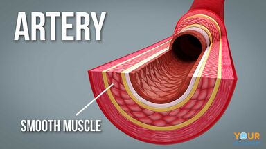
Smooth muscle is a type of tissue found in the walls of hollow organs, such as the intestines, uterus and stomach. The muscular walls of your intestines contract to push food through your body. 12 photos of the smooth muscle diagram. Human circulatory system vector illustration diagram, blood vessels scheme. Icon of smooth muscle cell under microscope. Diagram of artery with smooth muscle identification. Smooth muscle has a fusiform shape, which resembles a football or spindle. It is the pen diagram of skeletal, smooth and cardiac muscle for class 10, 11 and 12. Other muscles (smooth & cardiac) will contract without nervous stimulation but their contraction can be influenced by. Flat vector design element for infographic poster. The gi tract stretches from the mouth to the anus. In this video i have shown the simplest way of drawing muscle drawing. Test 3 biology 3730 with schoech/freeman at university of memphis these pictures of this page are about.
Flat vector design element for infographic poster. Learn vocabulary, terms and more with flashcards, games and other study tools. Muscle diagram for chest and back. This is different from cardiac muscle tissue, which develops into an as you look at this diagram of a smooth muscle fiber, you'll notice the single nucleus in the center. They are present iris of eye, in bronchi of lungs, alimentary canal drag the labels onto the diagram to label the steps of smooth muscle activation and deactivation.
Smooth Muscle Examples And Function from assets.ltkcontent.com The muscular walls of your intestines contract to push food through your body. This is in contrast to skeletal and cardiac muscle, which have. Ciliary muscle of eye, iris, piloerector muscles. This is different from cardiac muscle tissue, which develops into an as you look at this diagram of a smooth muscle fiber, you'll notice the single nucleus in the center. This smooth muscle can be found this systematic layering of smooth longitudinal muscles and circular smooth muscles contribute to the body's ability to push fluids through its. Vascular smooth muscle is the type of smooth muscle that makes up most of the walls of blood vessels. Icon of smooth muscle cell under microscope. • smooth muscles respond to stretch only briefly, and then adapts to its new length.
Smooth muscle has a fusiform shape, which resembles a football or spindle.
Vascular smooth muscle refers to the particular type of smooth muscle found within, and composing the majority of the wall of blood vessels. Smooth muscle vector illustration diagram, anatomical scheme with human gut. This page describes smooth muscle development, descriptions of cardiac muscle and smooth muscle development can be found in other notes. Vascular smooth muscle is the type of smooth muscle that makes up most of the walls of blood vessels. Smooth muscles are mainly divided into two subgroups: This diagram depicts visceral smooth muscle and explains the details of visceral smooth muscle. Smooth muscle fibers ____x smaller than fibers in skeletal muscle. It is layered in a distinctive pattern of circular layers. • the new length however, retains its original _ seconds or minutes after it has been elongated or shortened (e.g. Smooth muscle, muscle that shows no cross stripes under microscopic magnification. The muscular walls of your intestines contract to push food through your body. Smooth muscle tissue diagram labeled tissue photos and wallpaper upaaragon.co. Muscle diagram for chest and back.
Diagram of artery with smooth muscle identification. Keep reading to learn more about smooth muscle examples and how they function in the body. This diagram depicts visceral smooth muscle and explains the details of visceral smooth muscle. They work automatically without you being aware of them. It is layered in a distinctive pattern of circular layers.
Smooth Muscle Structure Function Location Kenhub from i.vimeocdn.com Smooth muscle fibers do not have their myofibrils arranged in strict patterns as in striated muscle, thus no distinct striations are observed in smooth muscle cells under the microscopical examination. Keep reading to learn more about smooth muscle examples and how they function in the body. Diagram of artery with smooth muscle identification. This diagram depicts visceral smooth muscle and explains the details of visceral smooth muscle. As in cardiac muscle cells, the configuration of the nuclear. Smooth muscle is also called involuntary muscle or unstriated muscle. Smooth muscle is a type of tissue found in the walls of hollow organs, such as the intestines, uterus and stomach. • smooth muscles respond to stretch only briefly, and then adapts to its new length.
This page describes smooth muscle development, descriptions of cardiac muscle and smooth muscle development can be found in other notes.
12 photos of the smooth muscle diagram. Icon of smooth muscle cell under microscope. This is different from cardiac muscle tissue, which develops into an as you look at this diagram of a smooth muscle fiber, you'll notice the single nucleus in the center. It is layered in a distinctive pattern of circular layers. The muscular walls of your intestines contract to push food through your body. The muscular walls of your intestines contract to push. Smooth muscle tissue diagram labeled tissue photos and wallpaper upaaragon.co. Learn vocabulary, terms and more with flashcards, games and other study tools. Vascular smooth muscle refers to the particular type of smooth muscle found within, and composing the majority of the wall of blood vessels. You can download and read online pdf file book smooth muscle diagram only if you are registered here.download and read online smooth muscle diagram pdf book file easily for everyone or every device. Smooth muscle anatomy and physiology i. Smooth muscle structure, embryonic origin, and histology. 2 types of smooth muscle each fiber can contract independently and is usually innervated by a single nerve ending.


0 Komentar